
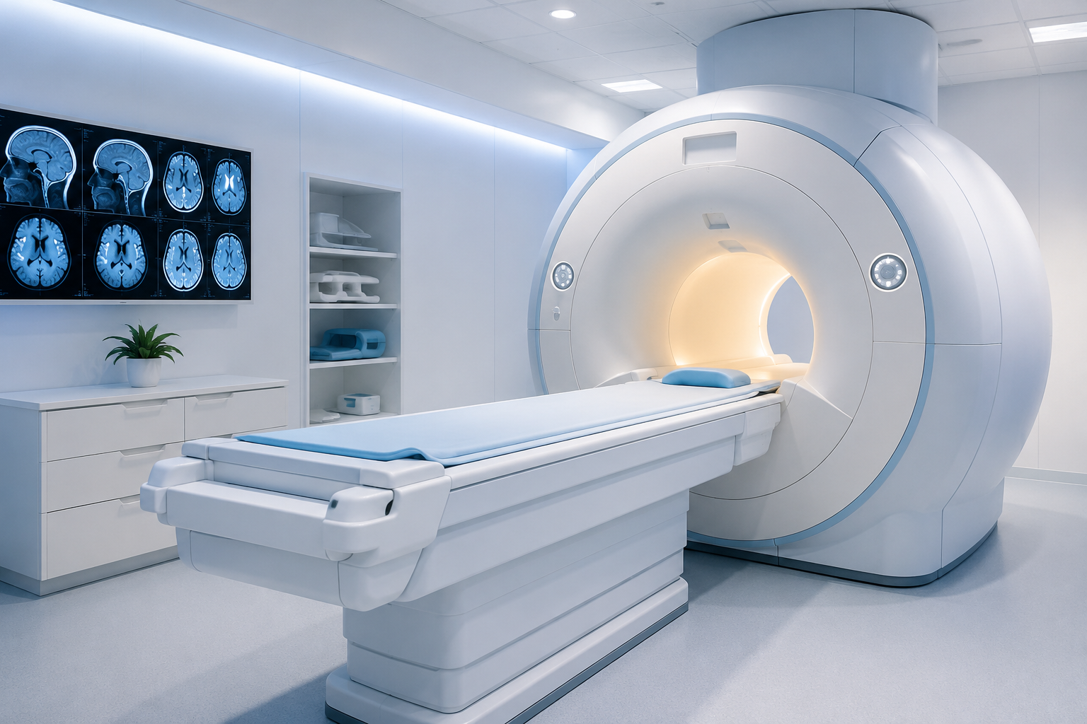
You received a referral for a brain MRI. Maybe it followed a headache your doctor wanted to investigate further. Maybe it came after a first seizure, a bout of dizziness, or a vision change. Whatever the reason, your first question is probably a simple one: Why?
A brain MRI is one of the most powerful diagnostic tools in modern medicine, capable of producing detailed images of brain tissue, blood vessels, and surrounding structures that no other imaging method can match. But the scan itself can feel opaque to patients who are not sure what it is looking for or what the results might mean.
This guide explains when and why doctors order brain MRIs, what the scan can detect, how it compares to a CT scan, and what Jersey City patients can expect from the process.
What Is a Brain MRI?
A brain MRI (magnetic resonance imaging scan of the brain) uses a powerful magnetic field and radio waves, not radiation, to create highly detailed cross-sectional images of the brain, brainstem, cranial nerves, blood vessels, and surrounding fluid-filled spaces.
Unlike an X-ray or CT scan, MRI does not use ionizing radiation, making it safe for repeated use and preferred for patients who need ongoing monitoring. It produces far greater soft-tissue contrast than CT, which means it can distinguish between different types of brain tissue, detect subtle lesions, and reveal abnormalities that may be invisible on other imaging modalities.
What the Machine Actually Captures
A brain MRI is not a single image; it is a series of sequences, each optimized to highlight different tissue properties. Depending on what your doctor has ordered, your scan may include:
- T1-weighted images: Show the brain’s anatomy in fine structural detail
- T2 and FLAIR sequences: Highlight fluid-filled areas, white matter changes, and lesions, particularly valuable for MS and small vessel disease
- Diffusion-weighted imaging (DWI): Detects acute stroke within minutes to hours of onset
- MR Angiography (MRA): Visualizes blood vessels to assess for aneurysms, stenosis, or vascular malformations
- Contrast-enhanced sequences: Gadolinium contrast is injected to highlight areas of blood-brain barrier breakdown, tumors, infection, or active inflammation
Important: A standard brain MRI typically takes 30–60 minutes. If contrast is ordered, add 15–20 minutes. Multiple sequence types are run during a single visit your doctor can specify which protocols are needed when ordering the scan.
Common Reasons Doctors Order a Brain MRI
A brain MRI referral is not a cause for alarm; it is a precision diagnostic tool that gives your doctor the clearest possible picture of what is happening inside your head. Here are the most common clinical reasons neurologists, primary care physicians, and specialists in Hudson County order brain MRIs.
1. Persistent, Severe, or New-Pattern Headaches
Most headaches are benign tension, migraine, or cluster headaches and do not require imaging. However, your doctor may order a brain MRI if your headaches present with certain warning signs:
- A new type of headache you have never experienced before
- Headache described as the “worst of your life” (thunderclap headache), a possible sign of aneurysm rupture
- Headaches that worsen progressively over weeks
- Headache accompanied by fever, stiff neck, visual changes, weakness, or confusion
- Headache that wakes you from sleep
- Headache following a head injury
For straightforward migraine or tension headache without neurological symptoms, brain MRI is generally not the first step. But when red flags are present, it is an essential tool for ruling out serious underlying causes.
2. First Seizure or Recurring Seizures
A brain MRI is a standard part of the evaluation for any new-onset seizure in an adult. The scan looks for structural causes such as cortical dysplasia, hippocampal sclerosis, tumors, or scarring from prior stroke that may be triggering seizure activity. For patients with known epilepsy, brain MRI helps guide treatment planning and monitor disease progression.
3. Neurological Symptoms: Numbness, Weakness, or Vision Changes
Sudden or progressive neurological symptoms numbness, weakness on one side of the body, arm or leg coordination problems, slurred speech, sudden vision changes can indicate a range of conditions, from stroke and transient ischemic attack (TIA) to multiple sclerosis, brain tumors, or other structural issues. Brain MRI, particularly with diffusion-weighted imaging, is the preferred scan for evaluating these symptoms.
4. Diagnosing or Monitoring Multiple Sclerosis (MS)
MRI is the gold standard for diagnosing multiple sclerosis. The scan reveals the characteristic white matter lesions (plaques) that MS produces in the brain and spinal cord, typically visible on FLAIR sequences. Brain MRI is also used regularly to monitor MS progression, assess response to treatment, and detect new or enlarging lesions before symptoms change.
5. Stroke Evaluation and Follow-Up
In the emergency setting, CT is typically the first scan performed because it is faster and more widely available. However, MRI, particularly DWI sequences, is more sensitive for detecting early ischemic stroke than CT, sometimes within minutes of onset. Brain MRI is routinely used for post-stroke follow-up to assess damage, monitor recovery, and identify risk factors for recurrence.
6. Suspected Brain Tumor or Abnormal Mass
When a brain tumor is suspected based on symptoms, a CT finding, or a referral from a specialist brain MRI with contrast is the definitive imaging modality. Gadolinium contrast causes abnormal tissue to “light up,” helping the radiologist characterize the size, location, blood supply, and tissue behavior of a lesion. MRI provides far more detail than CT for soft-tissue brain tumors.
7. Memory Loss, Cognitive Decline, or Dementia Evaluation
Brain MRI is often part of the diagnostic workup for new or progressive memory problems and cognitive decline. It can reveal brain atrophy patterns consistent with Alzheimer’s disease, evidence of multiple small strokes (vascular dementia), or other structural contributors to cognitive symptoms. While MRI alone cannot diagnose Alzheimer’s, it is an important tool for ruling out other causes and supporting clinical diagnosis.
8. Head Injury and Traumatic Brain Injury (TBI)
Following significant head trauma, CT is usually performed first in emergency settings because it quickly identifies life-threatening bleeds, skull fractures, and acute swelling. Brain MRI is used as a follow-up when CT findings are inconclusive, when symptoms persist, or when the physician needs to assess diffuse axonal injury, microbleeds, or more subtle structural changes that CT can miss.
9. Tinnitus, Vertigo, and Unexplained Dizziness
Persistent or severe dizziness, unexplained tinnitus (ringing in the ears), or sudden hearing loss may prompt a brain MRI particularly a dedicated inner ear MRI or MRI with contrast to rule out acoustic neuroma (a benign tumor on the hearing nerve), cerebellar disorders, or other posterior fossa pathology.
Symptoms, Concerns & What Brain MRI Can Help Evaluate
| Symptom or Concern | Conditions a Brain MRI Can Help Evaluate |
|---|---|
| Persistent or New-Pattern Headaches | Tumors, aneurysms, hydrocephalus, venous sinus thrombosis. |
| First-Ever or Recurring Seizures | Cortical dysplasia, hippocampal sclerosis, brain tumors, stroke-related scarring. |
| Sudden Numbness, Weakness, or Tingling | Stroke, transient ischemic attack (TIA), multiple sclerosis, brain or spinal cord lesions. |
| Vision or Speech Changes | Stroke, optic neuritis, multiple sclerosis, posterior fossa lesions. |
| Memory Loss or Cognitive Decline | Dementia, Alzheimer’s disease changes, small vessel disease, normal pressure hydrocephalus (NPH). |
| Severe Dizziness or Balance Problems | Cerebellar disorders, acoustic neuroma, inner ear abnormalities, brainstem conditions. |
| Head Injury or Trauma | Microbleeds, brain contusions, diffuse axonal injury, subdural collections. |
| Known or Suspected Multiple Sclerosis (MS) | White matter lesion burden, new demyelinating activity, disease progression. |
| Monitoring a Known Brain Condition | Tumor response to treatment, aneurysm surveillance, and post-surgical follow-up imaging. |
Brain MRI vs. CT Scan: Which Does Your Doctor Order and Why?
Patients are often confused about the difference between a brain MRI and a CT (computed tomography) scan of the head. Both produce detailed images of the brain, but they use different technology and are best suited to different clinical situations.
CT scan: Uses X-rays (ionizing radiation) to produce rapid cross-sectional images. CT is completed in 5–10 minutes and is ideal for emergency settings detecting acute bleeds, skull fractures, and large masses quickly. It is the first-line scan in trauma and stroke emergencies.
Brain MRI: Uses magnetic fields and radio waves (no radiation) and takes 30–60 minutes. Produces far greater soft-tissue contrast and detail. Preferred for evaluating tumors, MS, dementia, epilepsy, subtle stroke findings, and vascular abnormalities.
When Each Is Preferred
| Situation | Preferred Imaging |
|---|---|
| Suspected Tumor, Multiple Sclerosis (MS), or Complex Soft-Tissue Brain Condition | Brain MRI – Superior soft-tissue contrast and detailed visualization of brain structures. |
| Acute Emergency: Head Trauma or Suspected Brain Bleed (ER Setting) | Tomografía computarizada – Faster imaging and widely available for emergency diagnosis. |
| First or New-Onset Seizure (Non-Emergency) | Brain MRI – Better at identifying structural abnormalities that may trigger seizures. |
| Stroke Monitoring and Follow-Up | Brain MRI – Diffusion-weighted imaging (DWI) detects early stroke changes more accurately than CT. |
| Post-Surgical Surveillance for a Known Brain Tumor | Brain MRI with Contrast – Helps evaluate treatment response and detect recurrence. |
| Suspected Skull Fracture Following Trauma | Tomografía computarizada – Provides the clearest visualization of bone injuries and fractures. |
| Chronic Headaches with Neurological Symptoms | Brain MRI – Preferred for evaluating soft tissues, blood vessels, and neurological conditions. |
| Rapid Assessment of an Unconscious Patient in the Emergency Room | Tomografía computarizada – Speed is critical for immediate evaluation; MRI may be performed later if needed. |
What to Expect from a Brain MRI in Jersey City
Preparation
- For most brain MRIs, you can eat, drink, and take medications as normal.
- If your scan requires contrast, you may be asked to fast for 4–6 hours beforehand and will need to disclose any history of kidney disease (gadolinium is processed by the kidneys).
- Tell the imaging team about any metal implants, surgical clips, pacemakers, cochlear implants, or other medical devices essential safety screening for any MRI.
- Arrive 15–30 minutes early for paperwork and a safety screening questionnaire.
- Wear loose, metal-free clothing. You may be asked to change into a gown.
During the Scan
You will lie on a padded table that slides into the MRI scanner. A plastic head coil which looks like a cage around your head is placed around your head to improve signal quality. This is standard for brain MRIs and does not touch your skin.
The machine will make loud knocking and thumping sounds throughout the scan. This is normal and caused by magnetic field changes. Earplugs or headphones are always provided. Music is available at most Jersey City imaging centers.
Stay as still as possible. Even small movements can blur the images. The scan typically takes 30–60 minutes. If contrast is administered, a technologist will inject it through an IV partway through the scan.
After the Scan
There is no recovery time for a standard brain MRI. You can resume normal activities immediately. If contrast was used, drink plenty of fluids for the rest of the day.
A board-certified radiologist will review your images and send a written report to your referring physician typically within 24–48 hours at most Jersey City outpatient imaging centers. Your doctor will review the findings with you and discuss next steps.
Frequently Asked Questions
Not necessarily. Most headaches including tension and migraine do not require imaging. However, if your headache is new, unusually severe, the “worst of your life,” accompanied by neurological symptoms, or has changed significantly in pattern or frequency, your doctor may order a brain MRI to rule out structural causes. Always follow your physician’s clinical judgment.
Brain MRI provides significantly greater soft-tissue detail than CT. It can detect multiple sclerosis plaques, early or small strokes (with DWI sequences), subtle tumors, brain inflammation, small vascular malformations, diffuse axonal injury from trauma, and early dementia-related atrophy that a CT scan often misses. For non-emergency brain conditions, MRI is almost always the preferred diagnostic tool.
Brain MRI does not use ionizing radiation. It uses a powerful magnetic field and radio waves, which are not known to cause tissue damage. MRI is safe for most patients, including those who need repeated scans. Patients with certain metallic implants, pacemakers, or other devices should disclose these before any MRI the imaging team will verify safety. MRI is generally avoided in the first trimester of pregnancy unless medically necessary.
A standard brain MRI typically takes 30–60 minutes inside the scanner. With contrast, add 15–20 minutes. Your total visit including check-in, preparation, and check-out will usually be 60–90 minutes. Specialized protocols (MR angiography, functional MRI) may take longer.
After your scan, a board-certified radiologist reviews the images and sends a written report to your referring physician. At most outpatient imaging centers in Jersey City, this takes 24–48 hours. For urgent or STAT cases, same-day or next-morning reports are available. Your doctor will contact you to discuss the findings.

